Human Lower Body Diagram - Infographic Diagram Of Lower Half Human Skeleton Anatomy System Anterior View 3d Human Anatomy Medical Diagram Educational And Human Body Concept Isolated On White Background Stock Photo Download Image Now Istock : Bones of the pelvis and lower back.
Human Lower Body Diagram - Infographic Diagram Of Lower Half Human Skeleton Anatomy System Anterior View 3d Human Anatomy Medical Diagram Educational And Human Body Concept Isolated On White Background Stock Photo Download Image Now Istock : Bones of the pelvis and lower back.. Woman holding a blackboard with an illustration of the human digestive system drawn on it in chalk. Posted in diagrams scalenes muscles. The bones of the pelvis and lower back work together to support the body's weight, anchor the abdominal and hip muscles, and protect the delicate vital organs of the vertebral and abdominopelvic cavities. The vertebral column of the lower back includes the five lumbar vertebrae, the sacrum, and the coccyx. The sight of the classroom and my teacher's voice that said, c'mon class, open your science books, page 41, chapter 3, human body systems.
The sight of the classroom and my teacher's voice that said, c'mon class, open your science books, page 41, chapter 3, human body systems. Studying human anatomy became interesting, thanks to her. If you're looking for a speedy way to learn muscle anatomy, look no further than our anatomy crash courses. In all, there are believed to be 80 organs in your body, all serving different functions and uses. Superficial and deep anterior muscles of upper body
Leg Concise Medical Knowledge from cdn.lecturio.com In turn, the spinal cord relays essential information between the brain and the body. Learn anatomy as you browse our collection of colorful, large and clearly labeled human body diagrams. A) free body diagram for the block; Photo of human anatomy for fans of human anatomy 10358267. The myology of the lower limb is also particularly well represented in this atlas of anatomy, with multiple anatomical charts and diagrams: The scent of those textbooks; Studying human anatomy became interesting, thanks to her. The diaphragm forms the upper surface of the abdomen.
The calf muscle, on the back of the lower leg, is actually made up of two muscles:
Your body organs range from your brain, heart, liver, skin, lungs, kidneys, intestines, stomach, bladder, etc. Diagram lower back wiring diagram advance together the brain and spinal cord make up the central nervous system. The major muscles of the abdomen include the rectus. Glands in the human body explained with diagrams. B) free body diagram of point p; Bones of the pelvis and lower back. Two forces (lower part of figure below) 1) the weight w exerted by the earth on the box. Daniel nelson on june 5, 2018 8 comments ! Mental stress influences the flow of hormones and other fluids as well. Learn anatomy as you browse our collection of colorful, large and clearly labeled human body diagrams. Do you ever wonder what the major organs of the body are and. These muscles help the body bend at the waist. The spine anatomy is a complex structure.
Studying human anatomy became interesting, thanks to her. This diagram of the human body shows a range of organs that are important to human anatomy.they include the brain, heart, lungs, spleen, muscles. Daniel nelson on june 5, 2018 8 comments ! The pain body map is part of an update of content on the website by the michael g. The sight of the classroom and my teacher's voice that said, c'mon class, open your science books, page 41, chapter 3, human body systems.
Human Body Systems Overview Anatomy Functions Kenhub from thumbor.kenhub.com See human body diagram stock video clips. The scent of those textbooks; If you're looking for a speedy way to learn muscle anatomy, look no further than our anatomy crash courses. Muscle charts of the human body for your reference value these charts show the major superficial and deep muscles of the human body. Learn anatomy as you browse our collection of colorful, large and clearly labeled human body diagrams. Woman holding a blackboard with an illustration of the human digestive system drawn on it in chalk. The first diagram summarizes the different muscular compartments (fascial compartments) of the thigh and leg, and the different fascias (crural fascia, intermuscular septum, interosseous membrane, adductor canal, fascia lata) This diagram of the human body shows a range of organs that are important to human anatomy.they include the brain, heart, lungs, spleen, muscles.
This diagram depicts human body map of organs with parts and labels.
This diagram depicts human body map of organs with parts and labels. The gastrocnemius is the larger calf muscle, forming the bulge visible beneath the skin. We hope this picture human body artery diagram in detail can help you study and research. Woman holding a blackboard with an illustration of the human digestive system drawn on it in chalk. Learn here the cavities of the human body. Two forces (lower part of figure below) 1) the weight w exerted by the earth on the box. The muscles of the abdomen protect vital organs underneath and provide structure for the spine. Labeled illustration chart on white. At the level of the pelvic bones, the abdomen. The study of the human body involves anatomy, physiology, histology and. Superficial and deep anterior muscles of upper body Bones of the pelvis and lower back. The myology of the lower limb is also particularly well represented in this atlas of anatomy, with multiple anatomical charts and diagrams:
Muscle diagrams are a great way to get an overview of all of the muscles within a body region. Stomach, liver, intestine, bladder, lung, testicle, uterus, spine, pancreas, kidney, heart. The spine diagram shown below, consists of many bones or vertebrae,soft discs,the spinal cord, and spinal nerves. The gastrocnemius is the larger calf muscle, forming the bulge visible beneath the skin. Studying these is an ideal first step before moving onto the more advanced practices of muscle labeling and quizzes.
The Top Level Of The Tree Of Human Body Parts Thbp Thbp Consists Of 9 Download Scientific Diagram from www.researchgate.net 2) the tension force t ' 3 exerted by the string on the block. The muscles of the abdomen protect vital organs underneath and provide structure for the spine. The first diagram summarizes the different muscular compartments (fascial compartments) of the thigh and leg, and the different fascias (crural fascia, intermuscular septum, interosseous membrane, adductor canal, fascia lata) The bones of the pelvis and lower back work together to support the body's weight, anchor the abdominal and hip muscles, and protect the delicate vital organs of the vertebral and abdominopelvic cavities. The major muscles of the abdomen include the rectus. Find free pictures, photos, diagrams, images and information related to the human body right here at science kids. The vertebrae, which stack like spools of thread, support the back and protect the spinal cord. The pain body map is part of an update of content on the website by the michael g.
The gastrocnemius is the larger calf muscle, forming the bulge visible beneath the skin.
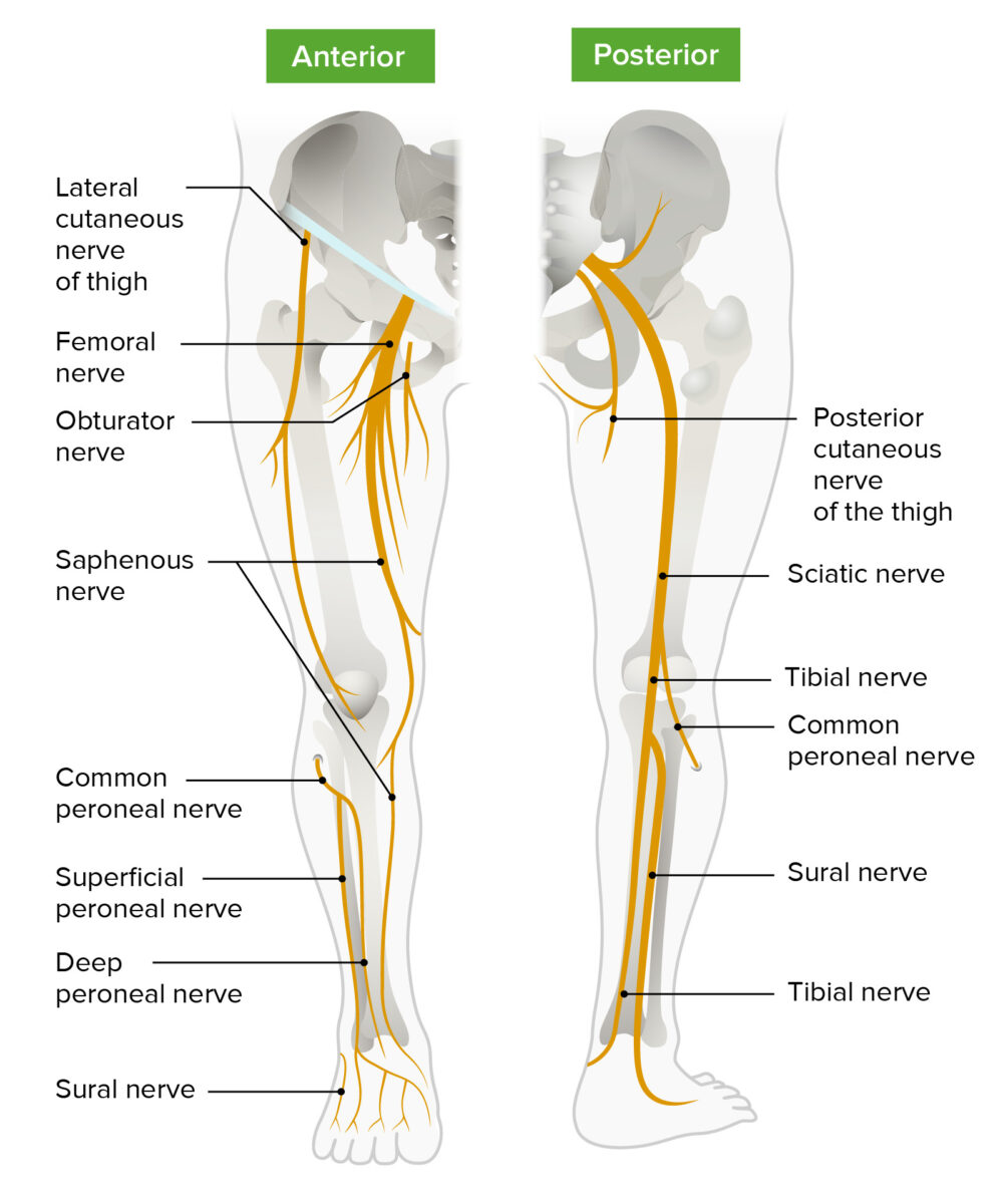
Find free pictures, photos, diagrams, images and information related to the human body right here at science kids. The vertebral column of the lower back includes the five lumbar vertebrae, the sacrum, and the coccyx. Labeled illustration chart on white. This diagram depicts human heart anatomy for kids 744×991 with parts and labels. We think this is the most useful anatomy picture that you need. The vertebrae, which stack like spools of thread, support the back and protect the spinal cord. These muscles help the body bend at the waist. See human body diagram stock video clips. In all, there are believed to be 80 organs in your body, all serving different functions and uses. The sciatic nerve is the dominant nerve that innervates the lower back and the lower extremities. 674 x 599 photo description: Riesige auswahl an cds, vinyl und mp3s. Anatomynote.com found human body artery diagram in detail from plenty of anatomical pictures on the internet.
We think this is the most useful anatomy picture that you need lower body diagram. The pain body map is part of an update of content on the website by the michael g.


0 Komentar